


Diagnostic Services
MRI, X-ray, Ultrasound & Specialist Scans
Rapid access through partner centres, 020 8501 0937
Book NowDiagnostic Imaging at Spine Plus
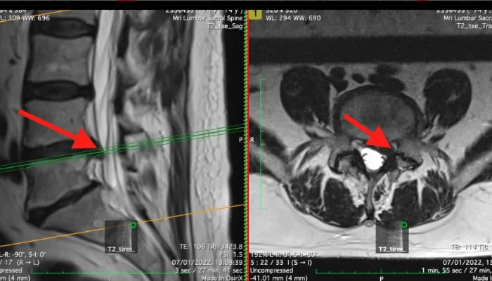
Accurate diagnosis is the foundation of effective treatment. Spine Plus provides rapid access to a full range of diagnostic imaging through our partner centres throughout London and Essex. When indicated, we can arrange scanning quickly, often within a few days.

Types of Scan Available
Standard MRI
Uses magnets and radio waves to produce detailed slice-by-slice images of the body. Shows bone, nerve, disc and ligament. Takes approximately 30 minutes. The investigation of choice for disc injuries, nerve problems, soft tissue pathology and spinal stenosis.
Open MRI
For patients who are claustrophobic. Uses an open bed rather than a tunnel. Image quality is slightly lower than standard MRI but suitable for most diagnostic purposes.
Seated / Upright MRI
Performed with the patient sitting or standing under load. Reveals disc problems that are not visible when the spine is unloaded in a lying position. Particularly useful for disc bulges that compress nerves only when weight-bearing.
3T MRI
Higher resolution than older 1.5T scanners. Better for detecting subtle pathology including small disc herniations, nerve root irritation and early degenerative changes.
Standard X-ray
Quick and cost-effective. Shows bone tissue. Identifies fractures, bone spurs, infections, tumours and alignment problems. Does not show soft tissue structures such as discs or ligaments.
Dynamic (Functional) X-ray
X-rays taken in sitting, standing, forward and backward bending positions. Reveals foraminal volumes, scoliosis, instability, facet joint changes and pelvic tilt. Information not visible on standard lying-down X-ray or MRI.
Ultrasound
Real-time imaging of muscles, tendons and ligaments near the body surface. Useful for peripheral joint problems (shoulder, elbow, wrist, knee, ankle). Limited use for deep spinal structures. Can also be used to guide injections.
SPECT/CT
Combines a radioactive tracer that identifies metabolically active "hot spots" with a low-field CT scan. Identifies pain-generating structures not visible on MRI or X-ray. Leads to more precise therapeutic intervention for complex or multi-level spinal conditions.
Other Tests
Nerve conduction tests and blood tests may be arranged where necessary to differentiate between conditions. We have access to the full range of investigations through our partner network.