


Diagnostics
MRI Scans, X-rays, Ultrasound & More
Through partner centres across London & Essex, 020 8501 0937
Book Now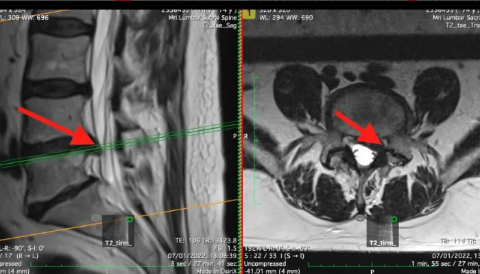
Imaging such as MRI and X-rays is performed at our various partner centres throughout London and Essex. Spine Plus provides rapid access to a full range of diagnostic services to support accurate diagnosis and treatment planning.
MRI Scans
Standard MRI Scan
Magnetic Resonance Imaging (MRI) uses magnets and radio waves to construct slice-by-slice computer images of the inside of the body. The images show all the relevant tissues including bone, nerve, disc and ligament. On average, these scans take around half an hour and involve lying still inside a scanner.
Open MRI Scan
Some patients find lying in the tunnel of a standard MRI scan claustrophobic. Open MRI machines do not feature a tunnel but use an open bed similar to an X-ray machine, making them more comfortable for patients who experience anxiety in enclosed spaces.
Seated / Upright MRI Scan
Upright MRI scans are performed with the patient sitting or standing rather than lying down. This can reveal problems such as disc bulging that is not evident when the spine is not under the loading of body weight, as is the case with standard lying-down MRI scans.
3T MRI Scan
The latest MRI scanners feature "3T" scanning, which gives a significantly higher image resolution than older 1.5T scanners. This improves the detection of subtle disc, nerve and ligament pathology.
X-rays
Standard X-ray
X-rays are quick and relatively cheap to perform. They do involve a small amount of radiation and primarily show bone tissue. Their main purpose is to identify bone fractures, infections, bone spurs (osteophytes), bone tumours and bony alignment problems. A normal X-ray does not rule out problems with non-bony structures such as discs, ligaments and nerves.
Dynamic (Functional) X-ray
Rather than taking an X-ray lying down, dynamic functional X-rays are performed with the patient sitting, standing, bending forward and bending backwards. This can reveal valuable information about foraminal volumes, scoliosis, facet joint hypertrophy, gas in the disc, degree of anterior listhesis, retrolisthesis and pelvic tilt. This information may not be visible on standard lying-down X-ray or MRI scans.
Ultrasound
Ultrasound scans are very useful for assessing damage in muscles, ligaments and tendons that lie near the surface. This is mostly used for problems in peripheral joints such as the shoulder, elbow, wrist, knee and ankle. Ultrasound has limited use for deep spinal problems.
SPECT/CT
Single Photon Computed Tomography (SPECT) uses a radioactive tracer injected into the bloodstream, which attaches to areas of unusual metabolic activity ("hot spots") that may indicate pain-generating sites not identified on MRI or X-ray. When combined with a low-field CT scan, better imaging of the pain-generating area is achieved so that specific structures (such as ligament, joint capsule or disc) can be identified as the source of pain. This leads to more precise therapeutic intervention.
Other Tests
It is sometimes necessary to refer patients for nerve conduction tests or blood tests to fully differentiate between certain conditions. At Spine Plus, we have access to the full range of such tests to get to the bottom of your problem.