



Spine Plus Blog
Modic Type 1 and Disc End Plate Changes
Published: 25 February 2024
What Are Modic Type 1 Changes?
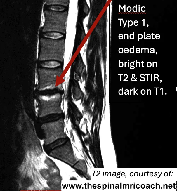
Modic changes are alterations in the vertebral bone marrow observed on MRI. Modic Type 1 changes specifically refer to regions of decreased signal intensity (dark) on T1-weighted images and increased signal intensity (bright) on T2-weighted images.
Since fat also produces hyperintensity on T2, a bright signal in the vertebra either side of a disc indicates either water or fat. A fat suppression sequence such as a STIR is the best type of sequence to definitively highlight Modic Type 1 changes.
In summary, Modic Type 1 bone marrow changes appear:
- Dark on T1
- Bright on T2
- Bright on STIR
This typically indicates the presence of oedema or inflammation within the vertebral body, and possibly minute stress fractures within the cancellous bone. In the context of disc herniation, Modic Type 1 changes often manifest adjacent to the affected intervertebral disc and are associated with degeneration of the cartilaginous end plate.
Causes of Modic Type 1 Changes
Modic changes are commonly associated with degenerative disc disease and are believed to result from a combination of mechanical, biological, and genetic factors:
- Microbial infection: Some studies suggest a potential link between Modic Type 1 changes and low-grade disc infections.
- Inflammatory response: Inflammation associated with end plate damage, possibly triggered by leakage of disc material into the vertebral body, leading to vascular changes and subsequent oedema.
- Mechanical stress: Chronic mechanical stress and instability in the affected spinal segment contributes to disc and end plate degeneration.
Modic Type 1 Changes in Disc Herniation
When a patient presents with a herniated disc, Modic Type 1 changes may be present due to the inflammatory response associated with disc end plate degeneration. The proximity of the inflammatory changes to the disc herniation suggests a dynamic relationship between disc pathology and vertebral bone alterations.
IDD Therapy and Modic Type 1 Changes
IDD Therapy, utilising distraction forces, has shown effectiveness in treating disc herniation. However, when Modic Type 1 changes are present, a more cautious approach is warranted.
Lighter Forces Initially
The use of lighter distraction forces is essential initially to avoid exacerbating pain associated with Modic Type 1 changes. The inflammatory nature of these changes may make the patient more sensitive to mechanical stresses. Gradual progression in force application allows for adaptation and minimises the risk of triggering an undesirable inflammatory response.
Starting Tension
Where Modic Type 1 changes are present, consider starting tension at half body weight minus 30 lbs (HBW minus 30). Progression of tensions should be very gradual, subject to patient response.
Monitoring Patient Response
Regular assessment of the patient's response is crucial. If the patient experiences heightened pain or intolerance, reducing the distraction force or modifying the treatment frequency may be necessary.
Multidisciplinary Approach
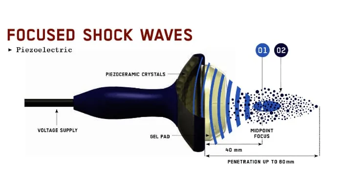
Collaborative communication between the treating clinician, radiologist, and other healthcare providers is vital. Additional modalities such as anti-inflammatory medications, focused shockwave, or TECAR therapy may complement IDD Therapy in addressing both the disc herniation and the associated Modic changes.
Conclusion
The presence of Modic Type 1 changes in patients with disc herniation warrants a tailored approach during IDD Therapy. By employing lighter forces initially and closely monitoring patient response, healthcare providers can strike a balance between effective treatment and minimising the risk of exacerbating symptoms. A collaborative, multidisciplinary approach ensures comprehensive care.