


Spine Plus Blog
MRI Points the Way
Published: 13 February 2020
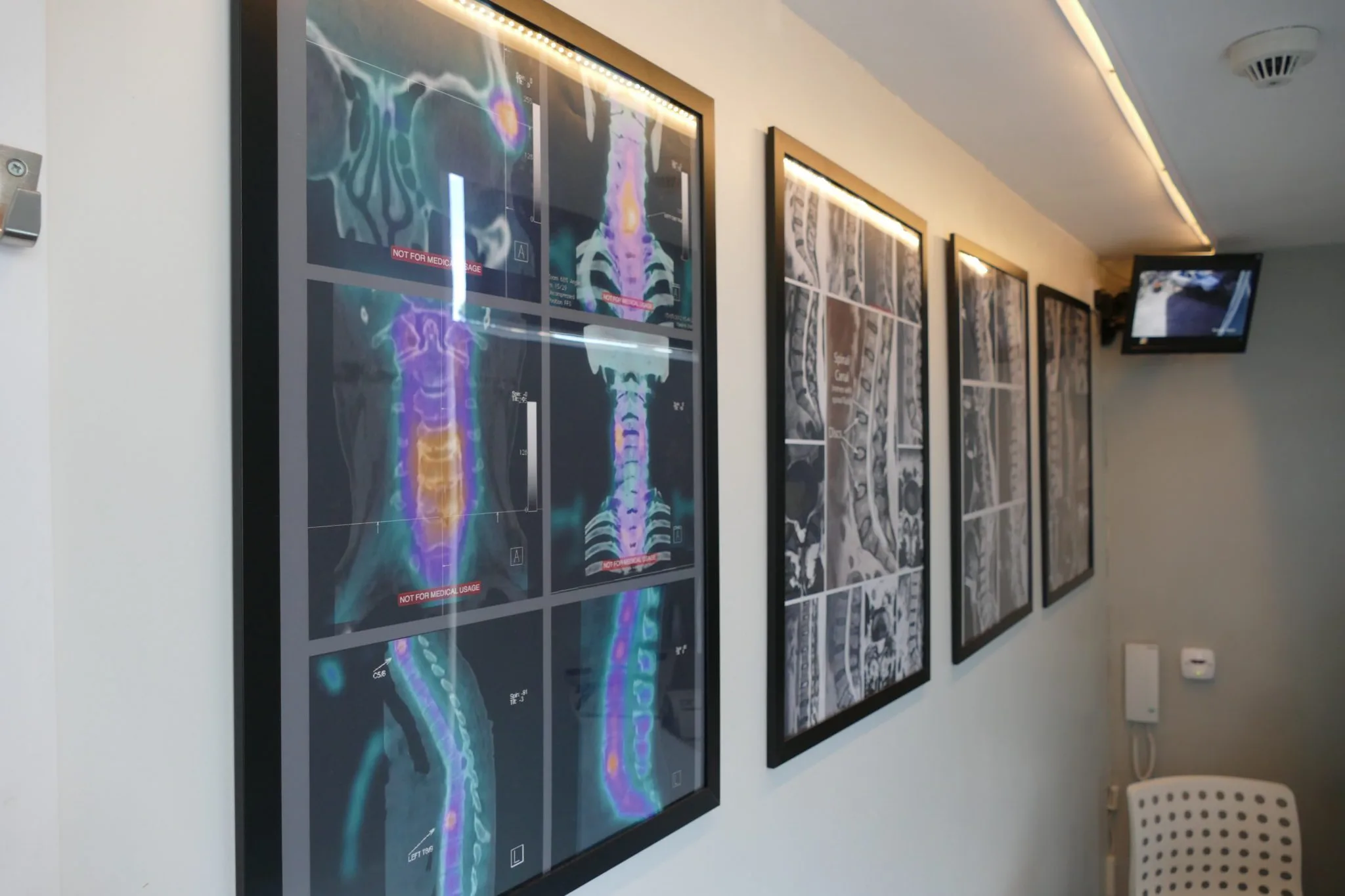
Jon came to us after years of chronic lower back pain. He'd seen multiple therapists who told him his hips and pelvis were misaligned. Spinal manipulation had become progressively less effective, and his back was "locking up" with increasing frequency.
What the MRI Showed
Clinical examination pointed to degenerative discs in the lower spine. An MRI confirmed: extensive Modic Type 1 change at L5-S1, facet osteoarthritis at the lower three motion segments, a left paracentral annular tear at L4-5, and a right paracentral annular tear at L5-S1. The imaging also showed slight instability of the L5 vertebra.
The findings matched the clinical picture: hypermobile L4/5 and L5/S1 segments with stiffness higher up, concentrating stress in the lower discs. This explained why manipulation had been progressively less useful, and more importantly, why it could have been making things worse by increasing motion at already unstable segments.
MRI-Led Treatment
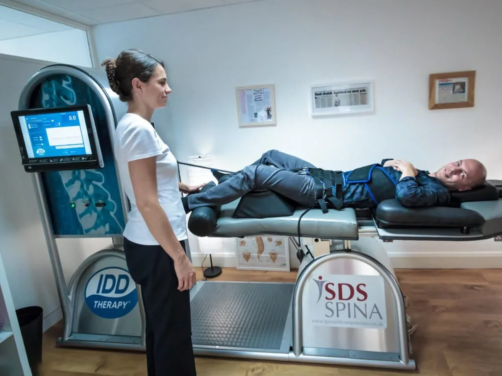
With clear imaging, treatment could be properly targeted. Spinal manipulation was stopped. The treatment programme included IDD Therapy for direct disc decompression, medical acupuncture to reduce muscle tension and spasm, core stabilisation exercises focused on the hypermobile lower segments, and prolotherapy administered by a sports doctor to strengthen the lax ligaments.
Outcome
After 12 to 24 months of consistent treatment and rehabilitation, Jon regained function and returned to playing football without back spasms. He avoided surgery.
If you have long-standing back pain with an unclear cause, an MRI is often the key to unlocking the right treatment. Call us on 020 8506 1000 to discuss a referral.