

Spine Plus Blog
Transitional Lumbosacral Joints
Published: 14 November 2019
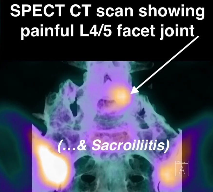
Studies indicate that approximately one third of people have a transitional lumbosacral joint, involving either lumbarisation of S1 (creating an extra disc space) or sacralisation of L5 (fusing it to the sacrum). The fusion can vary in degree and is classified using the Castellvi system.
Why It Matters
Movement and disc health: Sacralisation of L5/S1 causes movement restriction and protection of the L5/S1 disc, but shifts stress upward and accelerates degeneration at L4/L5. Many patients with a "bad" L4/L5 disc have sacralisation as an underlying factor that's never been identified.
Pseudo-arthrosis: Types 2 and 4 partial fusions are linked to increased low back pain severity and osteoarthritic changes in the pseudo-joint. This can require manual therapy alongside interventional procedures such as injections or radiofrequency treatment.
Neurological variations: Patients often have atypical nerve root distributions that don't match standard anatomical expectations. This can make clinical diagnosis confusing, as the pain pattern doesn't follow the expected dermatome.
Far Out syndrome: The pseudo-arthrosis can create bony or soft tissue lesions compressing nerve roots at the exit foramen. These are sometimes only visible on coronal MRI views that budget imaging providers may omit.
Clinical Relevance
Manual therapists likely encounter transitional lumbosacral joints frequently, whether or not they're specifically recognised. Proper identification through MRI or X-ray allows treatment to be tailored appropriately rather than applying a standard approach that may not address the underlying anatomy.
Call us on 020 8506 1000 to discuss an MRI assessment and diagnosis.